Site intended for healthcare professionals. Visit patient
site.

How 
Micro-Ultrasound works


 No area of concern observed on ultrasound.
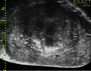
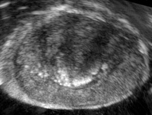
 Concerning area with mottled appearance observed on micro-ultrasound.
| Confirmed Gleason Grade Group 3 (4+3) PCa (80% pattern 4 with extensive cribriforming) |